
This article was written by Dr. Elena Borrelli, a Doctor of Medical Science and Physician Associate. Dr. Borrelli practices both clinical and academic medicine. She has over 20 years of experience working in healthcare, specifically in the following specialties: surgery, urgent care medicine, and ENT. Dr. Borrelli has authored several articles and other publications. She has also worked as a medical consultant and subject matter expert on various projects.
Nose Bleeds
Have you ever wondered why the nose bleeds so easily? Nosebleeds, also known as epistaxis, are very common and can occur very quickly for a variety of underlying reasons. Nosebleeds can occur spontaneously or secondary to trauma. It is common for a person to wake up with blood on a pillow from a nosebleed at night, especially in a dry environment. Sometimes a small bump in the nose or a sneeze can trigger a nosebleed. Often when the nose bleeds, it bleeds and continues to bleed. It is essential to search for the underlying cause of a nosebleed to help treat the current nosebleed and prevent further episodes. So why does the nose bleed so quickly and so much? Understanding the anatomy of the nose and nasal cavity can help better understand the pathophysiology behind nosebleeds and answer the questions above.
Understanding Nose Anatomy
Nasal Cavity
The internal nasal cavity is divided into a right and left nasal cavity. The nasal vestibule is the area surrounding the external opening of the nasal cavity. The nasal septum is a midline structure that separates the right and left nasal cavities. Oftentimes, a person is found to have a deviated nasal septum, causing one side of the nasal cavity to be smaller compared to the other. This can lead to various symptoms, including nasal dryness, breathing problems, or nosebleeds.
Turbinates
Inside the nasal cavity are turbinates, small bony structures that help humidify and filter incoming air. The nasal turbinates include the inferior and middle turbinates, as well as the superior turbinate. If turbinates become irritated, they can swell, causing turbinate hypertrophy. Enlarged turbinates can lead to breathing problems as well as nosebleeds.
Meatus
The spaces beneath each turbinate are called nasal meatuses. The nasal sinuses drain into the nasal meatuses.
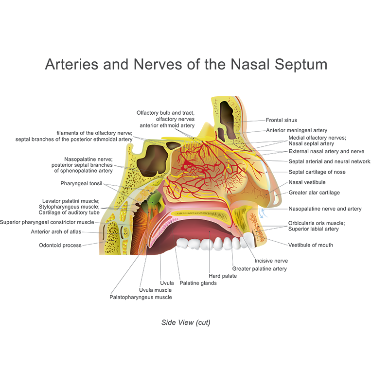
Vasculature
The nose contains a large number of blood vessels that join together to form different vascular plexus or networks. The extensive vascularity of the nose plays a large role in the cause of epistaxis.
Anterior Nosebleeds
Nosebleeds can be divided into anterior nosebleeds and posterior nosebleeds. Anterior nosebleeds are far more common and are often caused by bleeding from the Kiesselbach’s plexus. The Kiesselbach’s plexus is a collection of arteries that supply oxygen-rich blood to the nasal septum, which divides the nasal cavity into the right and left sides. The arteries that make up the Kiesselbach’s plexus include; anterior ethmoidal, greater palatine, sphenopalatine, and superior labial arteries. Due to the large number of arteries located all in one area, minor injury or irritation to the area can cause easy bleeding from the nasal septum. It is for this reason that the site of anterior nosebleeds is most commonly located on the anterior nasal septum. Typically, anterior nosebleeds will have blood flowing from one side of the nasal cavity.
Posterior nosebleeds
Posterior nosebleeds occur much less often than anterior nosebleeds; however, they are often more severe; as they can be more challenging to control; leading to increased blood loss. Posterior nosebleeds typically occur due to bleeding from the Woodruff plexus. Similar to the Kiesselbach’s plexus, Woodruff’s plexus is composed of a collection of blood vessels coming together. The Woodruff plexus is located in the posterior lateral wall of the inferior meatus of the nasal cavity. Specifically, the vessels involved in composing Woodruff’s plexus include the following; pharyngeal, posterior nasal, sphenopalatine, and posterior septal arteries. Posterior nosebleeds often cause blood to flow from bilateral nasal cavities and into the oropharynx.
Disturbance of Nasal Anatomy
Disturbance or alteration to the nasal anatomy can trigger nosebleeds. For example, turbinate hypertrophy can increase the dryness of the nasal cavity, lead to irritation, and as a result, cause epistaxis. In addition, injury to the nasal septum, such as septal perforation caused by cocaine use, can lead to epistaxis. Trauma to the nasal cavity, even minor trauma, can trigger a nosebleed. Digital trauma to the nose (nose picking) is a common cause of nosebleeds, especially among children.
Recurrent nosebleeds are especially concerning because an undiagnosed underlying condition should be considered. A thorough evaluation by a healthcare provider is essential to help identify an underlying cause if one exists. The nasal cavity requires special equipment for evaluation. A nasal speculum, along with a light source, is often indicated to view anterior structures of the nasal cavity. Further visualization can be performed using a nasoscope (also called a rhinoscope).
The first step in preventing recurrent nosebleeds is to identify where in the nasal cavity the bleeding is coming from and then identify the underlying cause. Once the underlying cause is identified, a management plan can be created to stop the bleeding and implement prevention measures to avoid future episodes of nosebleeds.
References